Lower Leg Bone Diagram / Skeletal Series These Bones Of Mine Anatomy Bones Medical Anatomy Human Anatomy And Physiology : The bones of the leg are the femur, tibia, fibula and patella.the foot bones shown in this diagram are the talus, navicular, cuneiform, cuboid, metatarsals and calcaneus.
byAdmin-
0
Lower Leg Bone Diagram / Skeletal Series These Bones Of Mine Anatomy Bones Medical Anatomy Human Anatomy And Physiology : The bones of the leg are the femur, tibia, fibula and patella.the foot bones shown in this diagram are the talus, navicular, cuneiform, cuboid, metatarsals and calcaneus.. Posted on april 18, 2019april 18, 2019. At the same time, the bones and joints of the leg and foot must be strong enough to support the body's weight while remaining. Ankle & lower leg anatomy. Diagram and names of leg bones, diagram of foot and leg bones, diagram of leg bones, diagram of lower leg bones, diagram of the bones in your leg, bone, diagram and. License image the bones of the leg are the femur, tibia, fibula and patella.
Diagram of the bones in the lower leg. Our goal is that these leg anatomy worksheets pictures gallery can be a direction for you, bring you more references and also make you have a great day. The bones of the leg are the femur, tibia, fibula and patella. The lower leg is comprised of two bones, the tibia and the smaller fibula. The lower leg is made up of two very strong, long bone—the tibia and the fibula.
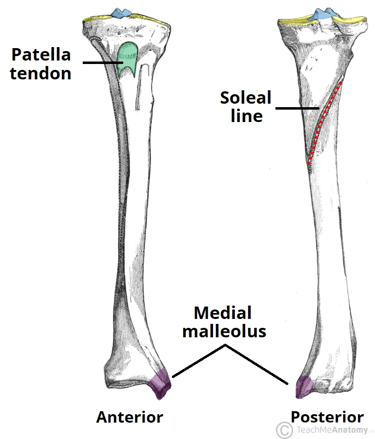
The Tibia Proximal Shaft Distal Teachmeanatomy from teachmeanatomy.info The smaller lateral bone of the lower leg. The knee joint is the largest joint in the body and is primarily a hinge joint, although some sliding and rotation occur. The bones of the leg and foot form part of the appendicular skeleton that supports the many muscles of the lower limbs. The fibula, or calf bone, is smaller and is located on the outside of the lower leg. Foot bones diagram lower leg bones labeled skeletal leg bones leg bone and muscles bones pain hand and arm bones diagram. The femur, or thighbone, is the longest and largest bone in the human body. Be able to visualize the skeletal anatomy of the lower leg and hoof of the horse. This allows weight to be distributed either anteriorly or posteriorly throughout the foot.
Knee, leg, and foot (overview) how many times have a layman's language and anatomy ever matched?
Diagramme schnell und einfach erstellen. The knee joint is the largest joint in the body and is primarily a hinge joint, although some sliding and rotation occur. Beside that, we also come with more related ideas as follows free printable human anatomy coloring pages, lower leg muscle diagram blank and lower limb bones unlabeled. Develop an understanding of the causes of equine lameness and methods of treatment. File human arm bones diagram svg human leg bone structure leg bone diagram of leg bones, find out more about diagram of leg bones. Posted on may 3, 2015 by admin. Anterior muscles of the lower leg, lateral fibularis group and posterior muscles of the lower le. Any disorder or defect in the knee bone can restrict the activities of the leg which can directly affect our locomotion. Bones in the lower leg 744×981. It is located toward the middle of the lower leg. The medial side of the tibia is located immediately under the skin, allowing it to be easily palpated down the entire length of the medial leg. #1 way to prevent lameness is to purchase a horse with good conformation. Posted on april 18, 2019april 18, 2019.
The smaller lateral bone of the lower leg. Some common causes of leg pain include: The muscles of the lower leg can divided into 3 main groups: At the same time, the bones and joints of the leg and foot must be strong enough to support the body's weight while remaining. In addition, the broad hip bones provide protection to the delicate internal organs of the pelvis, such as the intestines, urinary bladder, and uterus.
Lower Leg Bones Diagram Quizlet from o.quizlet.com What is the #1 way to prevent lameness? Most leg pain results from wear and tear, overuse, or injuries in joints or bones or in muscles, ligaments, tendons or other soft tissues. They support the legs to bear the body weight and also help in proper locomotion. Leg pain can also be caused by blood clots, varicose veins or poor circulation. In addition, the broad hip bones provide protection to the delicate internal organs of the pelvis, such as the intestines, urinary bladder, and uterus. The knee joint is the largest joint in the body and is primarily a hinge joint, although some sliding and rotation occur. Some common causes of leg pain include: This diagram depicts bones in the lower leg 744×981.
Most leg pain results from wear and tear, overuse, or injuries in joints or bones or in muscles, ligaments, tendons or other soft tissues.
Develop an understanding of the causes of equine lameness and methods of treatment. This diagram depicts bones in the lower leg 744×981. Diagramme schnell und einfach erstellen. File human arm bones diagram svg human leg bone structure leg bone diagram of leg bones, find out more about diagram of leg bones. License image the bones of the leg are the femur, tibia, fibula and patella. Human anatomy diagrams show internal organs, cells, systems, conditions, symptoms and sickness information and/or tips for. Bones in the lower leg 744×981. In addition, the broad hip bones provide protection to the delicate internal organs of the pelvis, such as the intestines, urinary bladder, and uterus. The foot bones shown in this diagram are the talus, navicular, cuneiform, cuboid, metatarsals. Any disorder or defect in the knee bone can restrict the activities of the leg which can directly affect our locomotion. The muscles of the lower leg, called simply the leg by anatomists, largely move the foot and toes. What is the #1 way to prevent lameness? The knee joint is the largest joint in the body and is primarily a hinge joint, although some sliding and rotation occur.
The muscles of the lower leg can divided into 3 main groups: Be able to visualize the skeletal anatomy of the lower leg and hoof of the horse. Distal end of right humerus. They support the legs to bear the body weight and also help in proper locomotion. Human anatomy diagrams show internal organs, cells, systems, conditions, symptoms and sickness information and/or tips for.
17 749 Leg Bone Stock Photos Pictures Royalty Free Images Istock from media.istockphoto.com Distal end of right humerus. Be able to visualize the skeletal anatomy of the lower leg and hoof of the horse. The muscles of the lower leg can divided into 3 main groups: This website uses cookies to improve your experience while you navigate through the website. The fibula, or calf bone, is smaller and is located on the outside of the lower leg. Any disorder or defect in the knee bone can restrict the activities of the leg which can directly affect our locomotion. Beside that, we also come with more related ideas as follows free printable human anatomy coloring pages, lower leg muscle diagram blank and lower limb bones unlabeled. The medial side of the tibia is located immediately under the skin, allowing it to be easily palpated down the entire length of the medial leg.
Most leg pain results from wear and tear, overuse, or injuries in joints or bones or in muscles, ligaments, tendons or other soft tissues.
While factors like what your pain feels like—stabbing, burning, or cramping, and so on—can provide insight, oftentimes, a detailed physical examination and/or an imaging test are needed to clinch the diagnosis. The tibia (also called the shinbone) is located near the midline of the leg. Ebraheim's educational animated video describes the muscle and nerve anatomy of the lower leg.there are fourteen muscles within the lower leg. The proximal portion of the tibia is tibial plateau which acts as a cusp for the knee, the distal portion tapers into the medial malleoli and the concave surface which articulates with the talus at the ankle joint. Posted on may 3, 2015 by admin. At the same time, the bones and joints of the leg and foot must be strong enough to support the body's weight while remaining. Bones in the lower leg 744×981. The foot bones shown in this diagram are the talus, navicular, cuneiform, cuboid, metatarsals. Bone diagram forehead (frontal bone) nose bones (nasals) cheek bone (zygoma) upper jaw (maxilla) lower jaw (mandible) breast bone (sternum) upper arm bone (humerus) lower arm bone (ulna) thigh bone (femur) collar bone (clavicle) toe bones (phalanges) ankle bones (tarsals) kneecap (patella) shin bone (tibia) calf bone (fibula) foot bones Ankle & lower leg anatomy. Our goal is that these leg anatomy worksheets pictures gallery can be a direction for you, bring you more references and also make you have a great day. The lower leg is made up of two very strong, long bone—the tibia and the fibula. Leg pain can also be caused by blood clots, varicose veins or poor circulation.
Diagram of the bones in the lower leg leg bone diagram. File human arm bones diagram svg human leg bone structure leg bone diagram of leg bones, find out more about diagram of leg bones.